This was the question at the forefront of my mind as I attended the Linz Winter Workshop back in February. Do biologists and physicists approach AFM differently and, does it matter?
The thought was triggered when I attended a stimulating talk given by ex-Bristol physicist Jamie Hobbs. Jamie is now a professor and biophysicist at the University of Sheffield and it was clear to me through his presentation that his grounding in physics is positively impacting the research.
Recently Jamie and the team were able to use AFM for the molecular imaging of glycan chains in e.coli.
As AFM provides high signal-to-noise imaging of individual biomolecules at the nanoscale, it has been considered a great tool to use in this area for a while. Being able to visualise the glycan chains is critical to understanding how the polymer network performs its function. However, despite considerable effort, successful imaging simply hasn’t been possible with AFM.
That is until Jamie and his team got to work on it. Guided by Jamie’s knowledge and confidence in using AFM they were able to develop new approaches to sample preparation and imaging which allowed glycans to be seen in high definition for the first time[1].
But what is the biologists view on working with AFM?
Not long after the conference I caught up with Daniel Robert, Professor of Bionanoscience at the University of Bristol to discuss the issue.

Professor Daniel Robert
(Image courtesy of University of Bristol)
Daniel highlighted that one of the challenges facing biologists is that AFM isn’t a core technique they study. Consequently, it often simply isn’t considered as a means of measurement or in terms of how it could be applied to biological research.
Indeed Daniel remembers that he first became aware of AFM during a talk giving by NuNano co-founder Mervyn Miles in the School of Biological Sciences back in 2002. The talk was on AFM of biomolecular structures, including the invention of the Activ Resonance controller[2], the first product of University of Bristol spin-out company Infinitesima.
Inspired by the talk and quick to see the connection between the work Merv had done and the research Daniel was undertaking on mosquitoes, he had a follow up chat with Merv. This resulted in Daniel using AFM to gain a deeper understanding of how mosquitoes hear.
Specifically, Daniel and team used AFM on live mosquitos to measure and characterise the mechanical response of mechanosensory neurons during forced oscillations of the insects hearing organs[3].
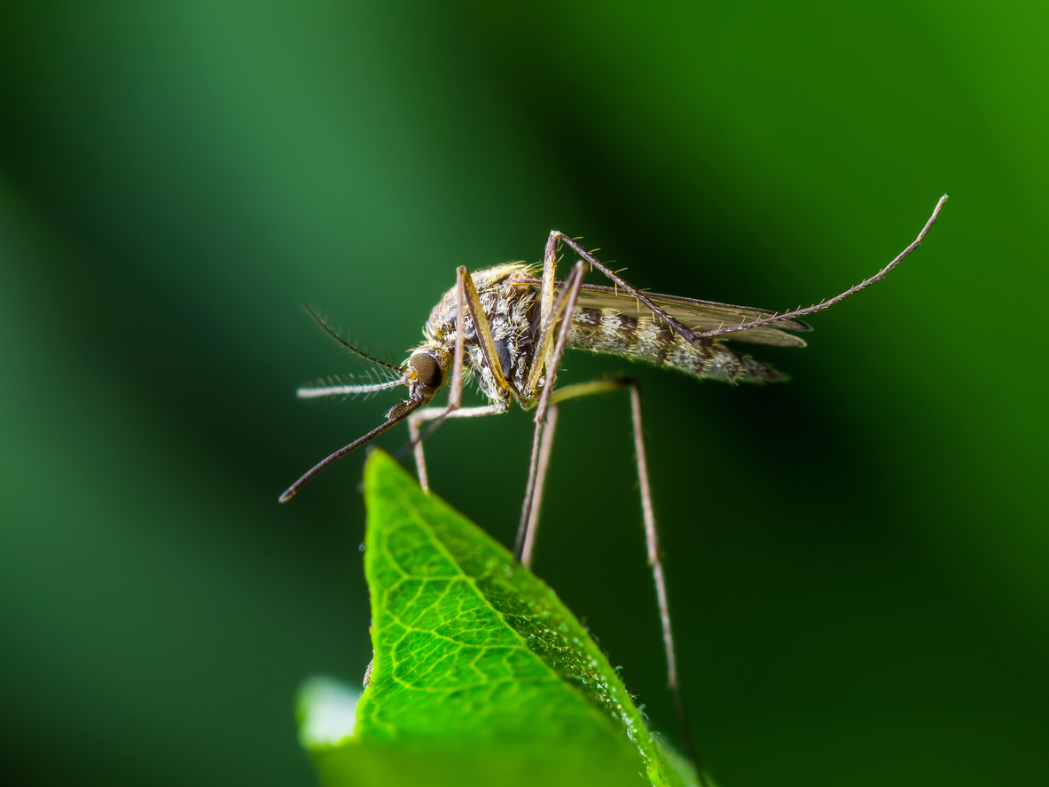
Everyone’s favourite insect…
Daniel and the team moved through different levels in using the AFM: From simple contact mode imaging, to more advanced measurements of the response and shape of the oscillation, as well as characterising the system’s linearity.
But the really new application the AFM enabled the team to undertake was to further their understanding on the role of electrostatics in sensory systems in insects, characterising whether the system was electric and how it acquired or dissipated charge.
For Daniel, realising that the AFM was a tool that could help him answer a biological question he was examining was a breakthrough. He agrees that there are fundamental differences between how a physicist and a biologist approach a problem but he believes that the best research involves both backgrounds. This is evident in the fact that Daniel ensures he always has a couple of physicists in the lab and on the papers he writes. He says:
“We don’t make the biologists physicists and we don’t make the physicists biologists. Somehow [working together] researchers emerge in yet another area – it’s not meeting in the middle between A and B, it’s meeting at C, somewhere else.”
This idea of ‘meeting at C’ as a third place is what I find really interesting. I find truly interdisciplinary working in this way energising and I’m fascinated by the novel developments occurring in the use of AFM through the questions posed by biology.
Coming back to my own, somewhat intentionally provocative question, yes, I think it’s fair to say biologists and physicists do approach AFM differently and, actually yes, it does matter. But not in a detrimental way to either.
Like Daniel, I think effective communication between physicists and biologists can transform a variety of research areas, aided by the application of any number of measurement techniques, such as AFM.
If you’re already using AFM in your current project, or thinking about it, we’re always keen to hear about it. We may even be able to offer you some key tips and advice on how to get the best out of your AFM and therefore produce even better results to aid your research.
[1] You can read more about this research in the article Molecular imaging of glycan chains couples cell wall polysaccharide architecture to bacterial cell morphology by Turner, Mesnage, Hobbs and Foster published in Nature Communications.
[2] The two papers that relate to the talk Merv was giving are: Piconewton regime dynamic force microscopy in liquid and High-Q dynamic force microscopy in liquid and its application to living cells.
[3] The resulting work on Frequency doubling by active in vivo motility of mechanosensory neurons in the mosquito ear was published in the Royal Society Open Science journal in January last year.
If you liked this blog post you might also like An interview with Professor Daniel Robert and The multidisciplinary nature of AFM
Need probes? Click here for our full range!